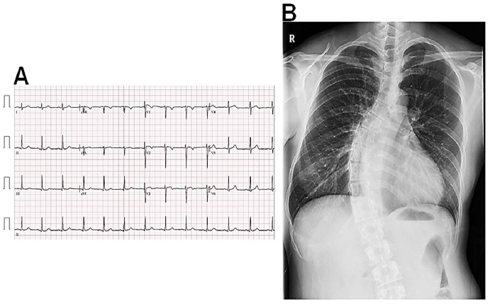
Figure 1: Electrocardiography and chest radiography at the initial admission Electrocardiography shows sinus rhythm with non-specific ST-T segment changes (A). Chest radiography shows mild cardiomegaly with tortuous thoracic spines (B).