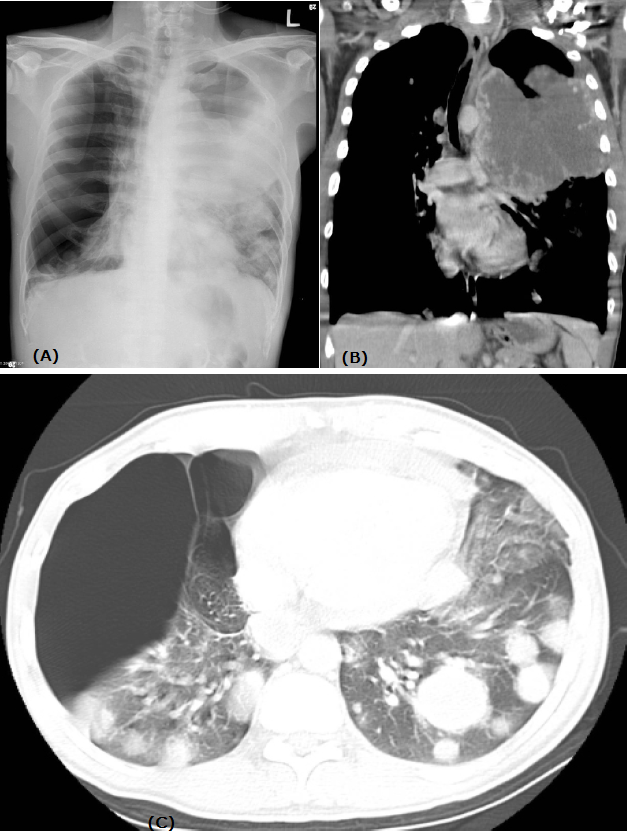
Figure 1: (A) Chest radiograph shows opacity of left lung and a hyperlucent right lung. There are multiple nodules in both lungs.
(B) Contrast-enhanced computed tomography (CT) reformatted image shows a bulky, ill-circumscribed mass with central necrosis arising from anterior mediastinum.
(C) Axial lung window setting shows multiple round nodules in bilateral lungs and large bullae in the right lung.
(B) Contrast-enhanced computed tomography (CT) reformatted image shows a bulky, ill-circumscribed mass with central necrosis arising from anterior mediastinum.
(C) Axial lung window setting shows multiple round nodules in bilateral lungs and large bullae in the right lung.