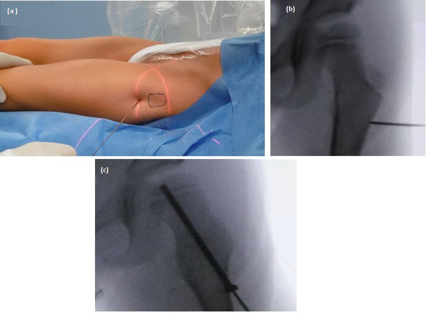
Figure 4: (a) Intraoperative situation, marking the entry point for the screw with the help of a wire. (b) Intraoperative fluoroscopy: wire approx. 2-3 cm below the greater trochanter. (c) Intraoperative fluoroscopic image: screw aligned in the axis of the femoral neck and good grip of the epiphyseal gap just slightly medial to the middle of the physis.