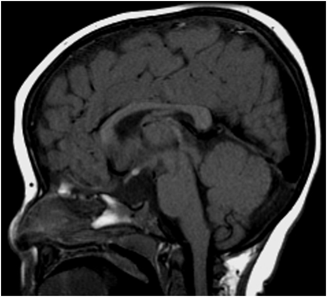
Figure 1:
Sagittal T1 weighted Magnetic resonance imaging showing a small anterior pituitary, absent stalk, and normally located posterior pituitary.