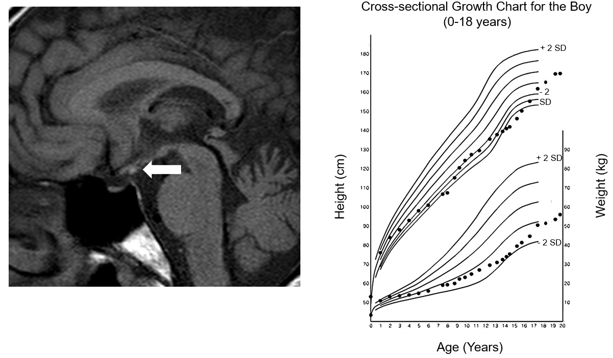
Figure 1: Magnetic resonance imaging findings and growth chart of the patient.
(A): T2-weighted magnetic resonance image of the pituitary region of the patient. The pituitary stalk could not be identified, and an ectopic posterior pituitary bright spot was noted (arrow). The size of the anterior pituitary gland revealed hypoplasia.
(B): Growth chart of the patient.
(A): T2-weighted magnetic resonance image of the pituitary region of the patient. The pituitary stalk could not be identified, and an ectopic posterior pituitary bright spot was noted (arrow). The size of the anterior pituitary gland revealed hypoplasia.
(B): Growth chart of the patient.