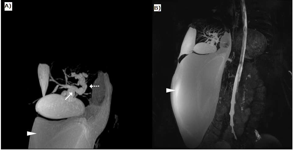
Figure 3: MRCP images. 3A: The proximal part of the common hepatic duct (arrow) and intrahepatic bile ducts are dilated. Multifocal strictures, dilatations and irregularity are seen, particularly in the left intrahepatic bile ducts (stippled arrow). There is a fluid collection suggesting biloma in the subhepatic region (arrowhead). 3B: MRCP image showing a huge loculated fluid collection extending from the subcapsular region of the right hepatic lobe to the pelvic inlet (arrowhead).