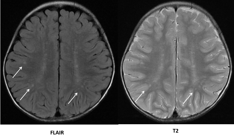
Figure 3: Fluid attenuated inversion recovery (FLAIR) and T2 weighted magnetic resonance imaging. High intensity areas on multiple white matters around the cerebral ventricles (white arrows) are shown in both FLAIR and T2 weighted images.