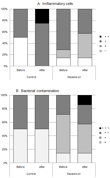
Figure 2: Cytological examination. Smears of oral mucosa were fixed, stained with Papanicolaou’s stain and examined by a qualified cytologist according to the following criteria:
- Inflammatory change: (- ) No inflammation; (±) very small number of inflammatory cells are observed; (+) Inflammatory cells are observed in less than one third of the fields: (++) Inflammatory cells are observed in more than one third of the fields; (+++ ) Severe inflammatory changes are observed in all fields.
- Bacterial contamination: (-)No bacterial cell are observed ; (±) very small number of bacterial cells are observed; (+) Bacterial cells are observed in several fields: (++)Bacterial cells without prominent colony formation are observed in entire background.; (+++ ) Bacterial cells with prominent colony formation are observed in entire background.