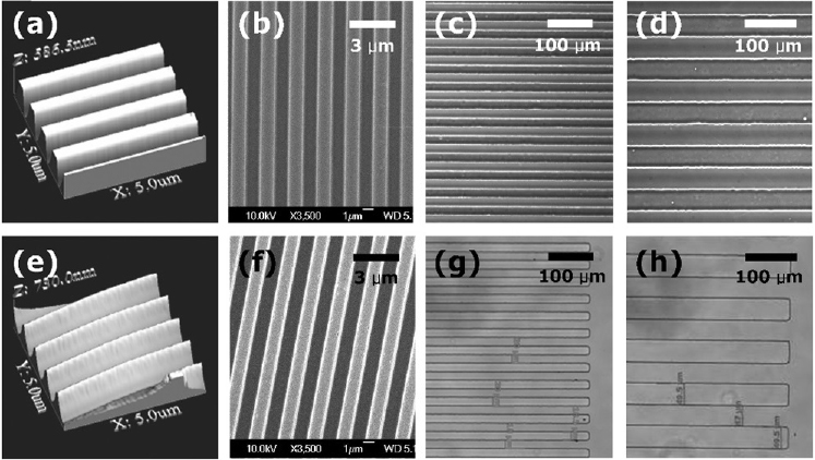
Figure 1: Various substrates for culture of MSCs. Various-dimensional groove and ridge patterns fabricated in SU-8 shown in (a)-(d), patterns fabricated in PDMS are shown in (e)-(h). (a) and (e) are the AFM (atomic force microscope) images of the substrates molded from a master with 500 nm × 500 nm × 500 nm (groove width × spacing width × depth) groove and ridge patterns. (b) and (f) are scanning electron microscopic images of the images1 μm × 1 μm × 1 μm. The others are microscopic images of 20 μm × 20 μm × 10 μm ((c) and (g)) and 50 μm × 50 μm × 10 μm ((d) and (h)) groove and ridge patterns.