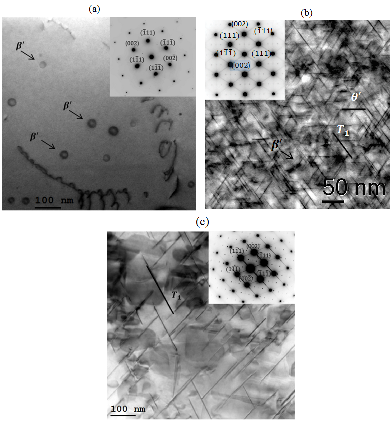
Figure 2: Evolution of the microstructure as observed by TEM.
(a) Typical bright field image of the specimen after conducting the T4 heat treatment shows the presence of β' precipitates within the microstructure and the corresponding [101] selected area diffraction pattern (SADP).
(b) Bright field image of the microstructure conducting T8 heat treatment condition shows a complex microstructure with the T1, θ' and β' precipitates and the corresponding [101] selected area diffraction pattern (SADP).
(c) Microstructure of the specimen after the T6 heat treatment condition consists of T1 precipitates as the primary strengthening phase and the corresponding [101] selected area diffraction pattern (SADP).