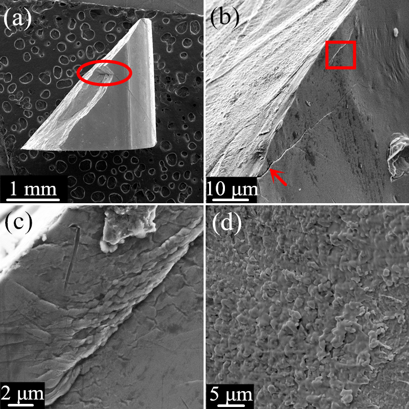
Figure 5: The fractographs of the composite upon dynamic compression. (a) The lateral surface of the whole deformed composite, from which, a piece of fragment, indicated by the red ellipse, has come away; (b) the magnified image for the lateral surface of the deformed sample, only primary shear bands and microcrack, indicated by the red arrow, can be seen; (c) the higher magnification of the area marked by the red rectangular in Figure 5(b), showing the details of the shear band; (d) the fracture surface of the composite.