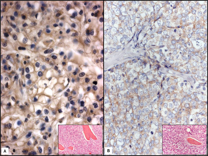
Figure 2: LF immunopositivity was well represented at the periphery of clear cell in a metastasis to bone by renal carcinoma (a, x400); the inset illustrated the routinely stained section with partially destroyed bone lamellae (x 40). A light to moderate LF immunostaining was found in breast ductal invasive carcinoma (b, x400); the inset illustrated the routinely stained section with partially destroyed bone lamellae (x 40). (Mayer's hemalum counterstain).