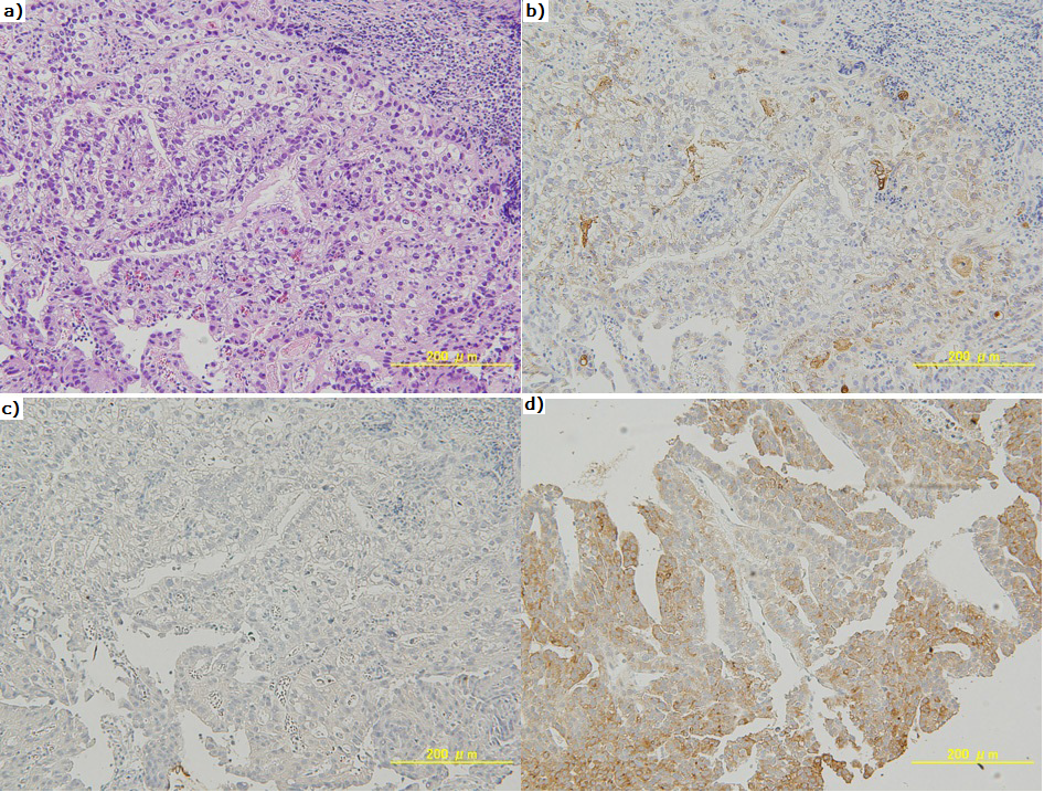
Figure 4: Gastric biopsy shows an adenocarcinoma. Cancer cell discloses irregular tubular and alveolar atypical epithelial cell including clear reticulum (a: hematoxylin and eosin stain, ×200). Immunohistochemical staining shows alpha-fetoprotein expression in the cancer cell (×200) (b), but no desgamma- carboxy prothrombin (DCP) expression (×200) (c). Histopathological findings of the liver tumor are similar to those of adenocarcinoma from the gastric biopsy specimen, although DCP is expressed in cancer cells (×200) (d).