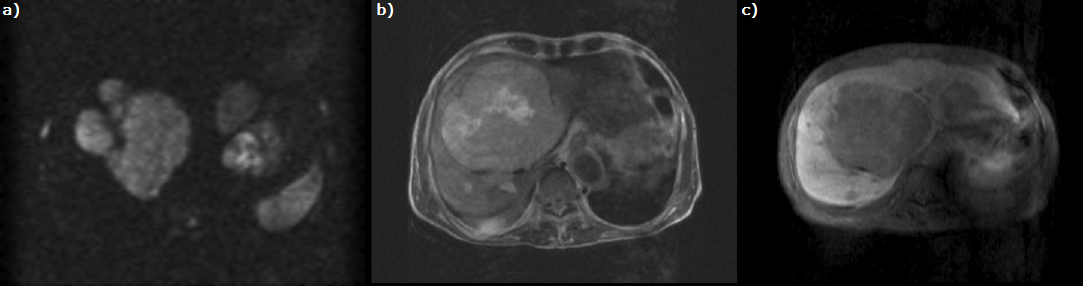
Figure 3: Contrast magnetic resonance imaging shows multiple nodules in the liver. The nodule inside has high intensity on T2- weighted imaging and diffusion-weighted imaging (a). The tumor is enhanced heterogeneously in the early phase and washed out in the late to hepatocellular phase (b, c).