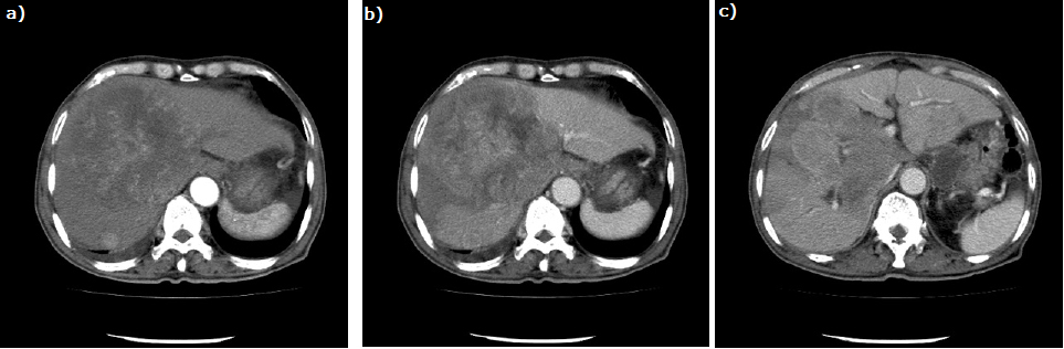
Figure 2: Abdominal computed tomography shows multiple low-density areas in the liver; the largest tumor is 13 cm in diameter. The tumor is enhanced at the peripheral zone and mosaic inside in the early phase and washed out in the late phase (a, b). A bulky gastric lymph node is also seen (arrow) (c).