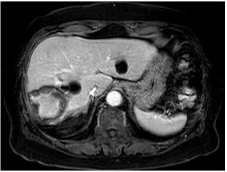
Figure 3:
Magnetic resonance imaging revealed a hypointense lesion with central hyperintensity on T1-weighted sequences (a) and hyperintensity on T2-weighted sequences (b).