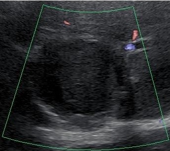
Figure 1: Ultrasonography revealed a protruding hypoechoic mass, 6 cm in diameter, with a central isoechoic structure in segment 7, accompanied by multiple hepatic cysts. On Doppler ultrasonography, no blood flow was detected in the hypoechoic mass.