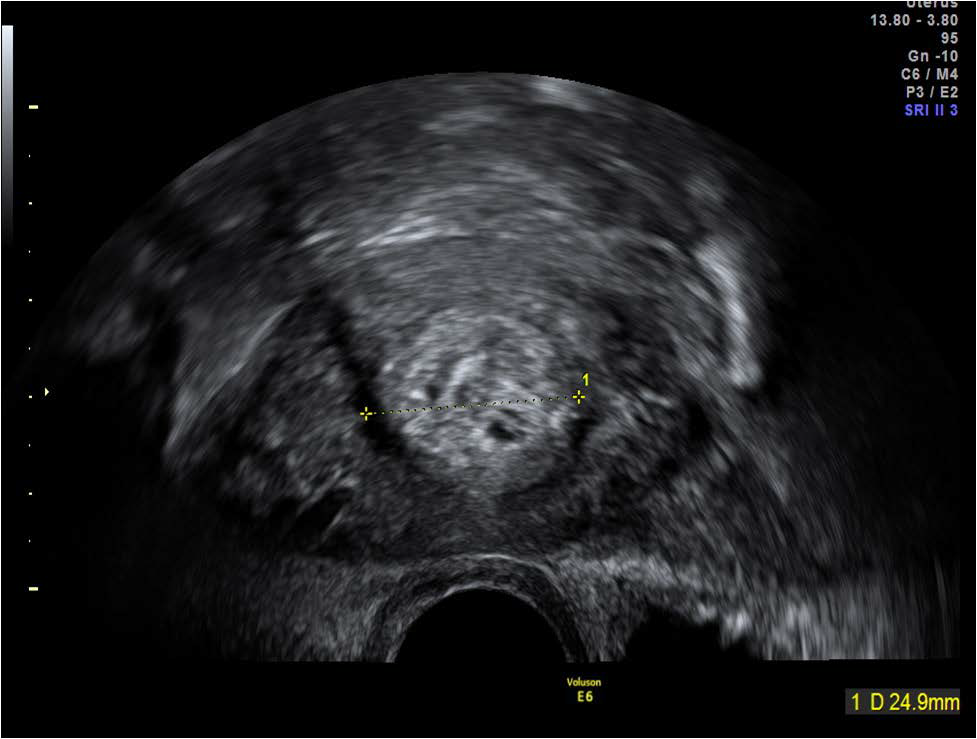
Figure 8:
Echogenic mass within the lower uterine segment in the same patient (Figure 7) as seen in the transverse plane on transvaginal scan.