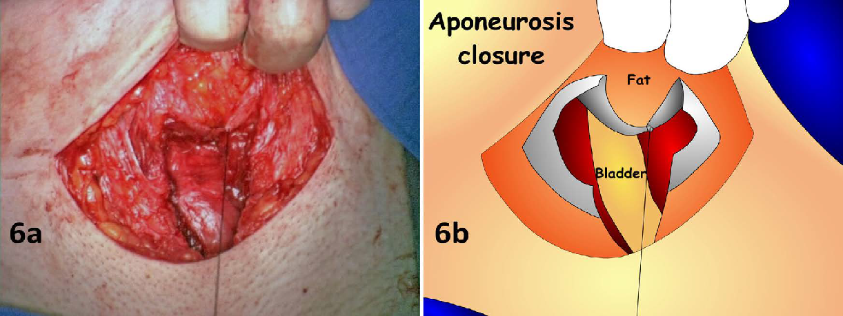
Figure 6: (6a) As fat tissues were not splitted from the aponeurosis, the aponeurosis closure begins with its posterior surface exposed. (6b) This scheme shows the everted upper edges to help visual control. The continuous suture starts in the upper corner and leads along the posterior surface of the anterior aponeurosis of the rectus abdominis muscle back to the initial incision point.