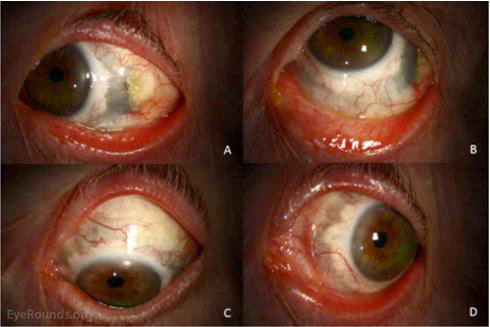
Figure 5: Anterior Necrotizing Scleritis at latest follow-up.
(Courtesy of Eye.Rounds.org University of Iowa)
The left eye is shown in right gaze (A), up gaze (B), down gaze (C), and left gaze (D). The conjunctival injection is markedly improved. The temporal conjunctival defect is beginning to epithelialize. Brown or dark pigmentation is now present from approximately 1 o'clock to 11 o'clock (clockwise). This represents direct visualization of the choroid and ciliary body due to loss of scleral tissue from the inflammatory process.