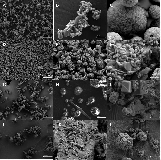
Figure 4:
Representative images of the HA particles obtained by scanning electron microscopy of the hydroxyapatite particles obtained by atomization: B1 (A, B, C); B2 (D, E, F); B3 (G, H, I); B4 (J, K, L).