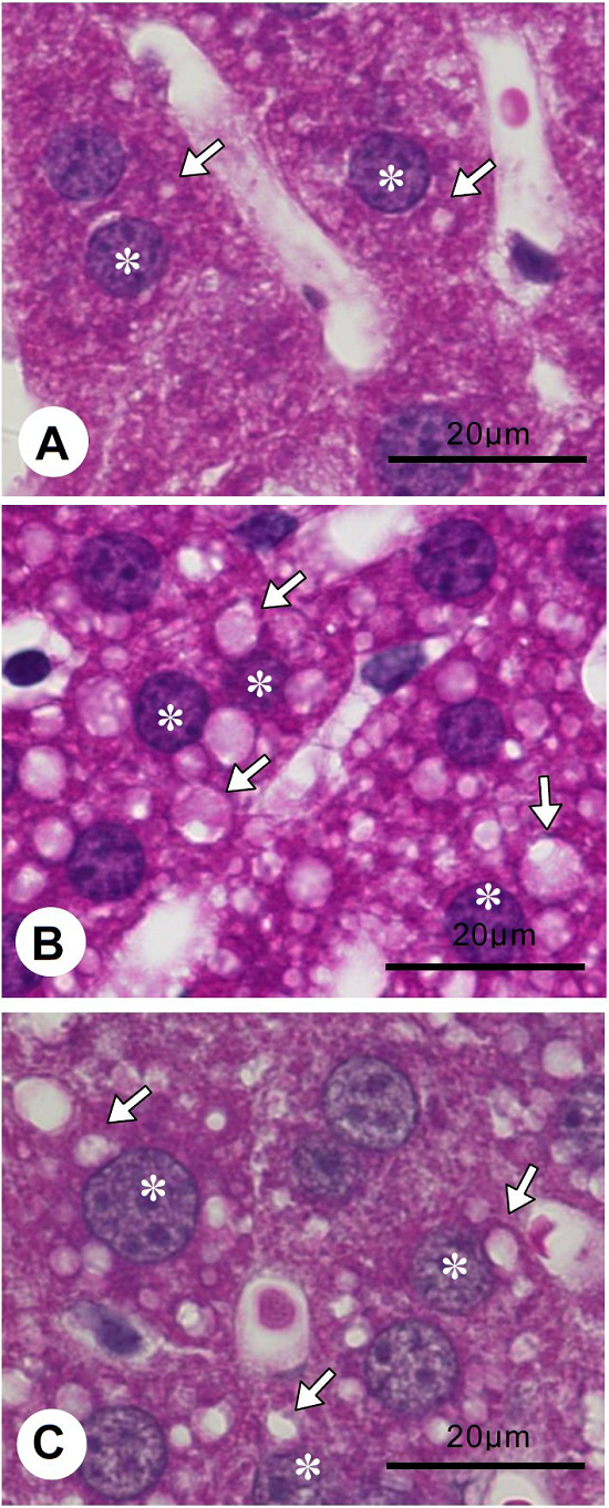
Figure 2: Comparison of the size and number liver lipid droplets (A)-(C) H&E staining of liver sections.
(A) Liver section from a normal mouse fed with control diet (CD). Lipid droplets (arrows) in hepatocytes (asterisk) are typically small in size (about 2 μm in diameter), and a couple of lipid droplets (2-3 pieces) are contained in a cell. SI: Sinusoid. (B) Liver section from a mouse fed with high fat-diet (HD). Lipid droplets (arrows) in hepatocytes (asterisk) are larger than that of normal liver (5-6 μm in diameter). Further, an increased number of droplets in cells (usually 5-7 per cell) is observed. SI: Sinusoid. (C) Liver section from a mouse fed with HD containing Lb. paracasei. The size of lipid droplets (arrows) in hepatocytes (asterisk) is smaller than that of animals fed HD (about 3 μm in diameter). The number of lipid droplets in each hepatocyte is also reduced (usually 2-3 per cell) in comparison to that of animals fed with HD.
(A) Liver section from a normal mouse fed with control diet (CD). Lipid droplets (arrows) in hepatocytes (asterisk) are typically small in size (about 2 μm in diameter), and a couple of lipid droplets (2-3 pieces) are contained in a cell. SI: Sinusoid. (B) Liver section from a mouse fed with high fat-diet (HD). Lipid droplets (arrows) in hepatocytes (asterisk) are larger than that of normal liver (5-6 μm in diameter). Further, an increased number of droplets in cells (usually 5-7 per cell) is observed. SI: Sinusoid. (C) Liver section from a mouse fed with HD containing Lb. paracasei. The size of lipid droplets (arrows) in hepatocytes (asterisk) is smaller than that of animals fed HD (about 3 μm in diameter). The number of lipid droplets in each hepatocyte is also reduced (usually 2-3 per cell) in comparison to that of animals fed with HD.